Currently, there is a need for noninvasive detection technologies that are able to assess skin barrier integrity and function that are both accurate and convenient. Several techniques and tools exist on the market, including laser imaging methods and Raman spectroscopy, however, dermatologic experts continue to search for a better solution.
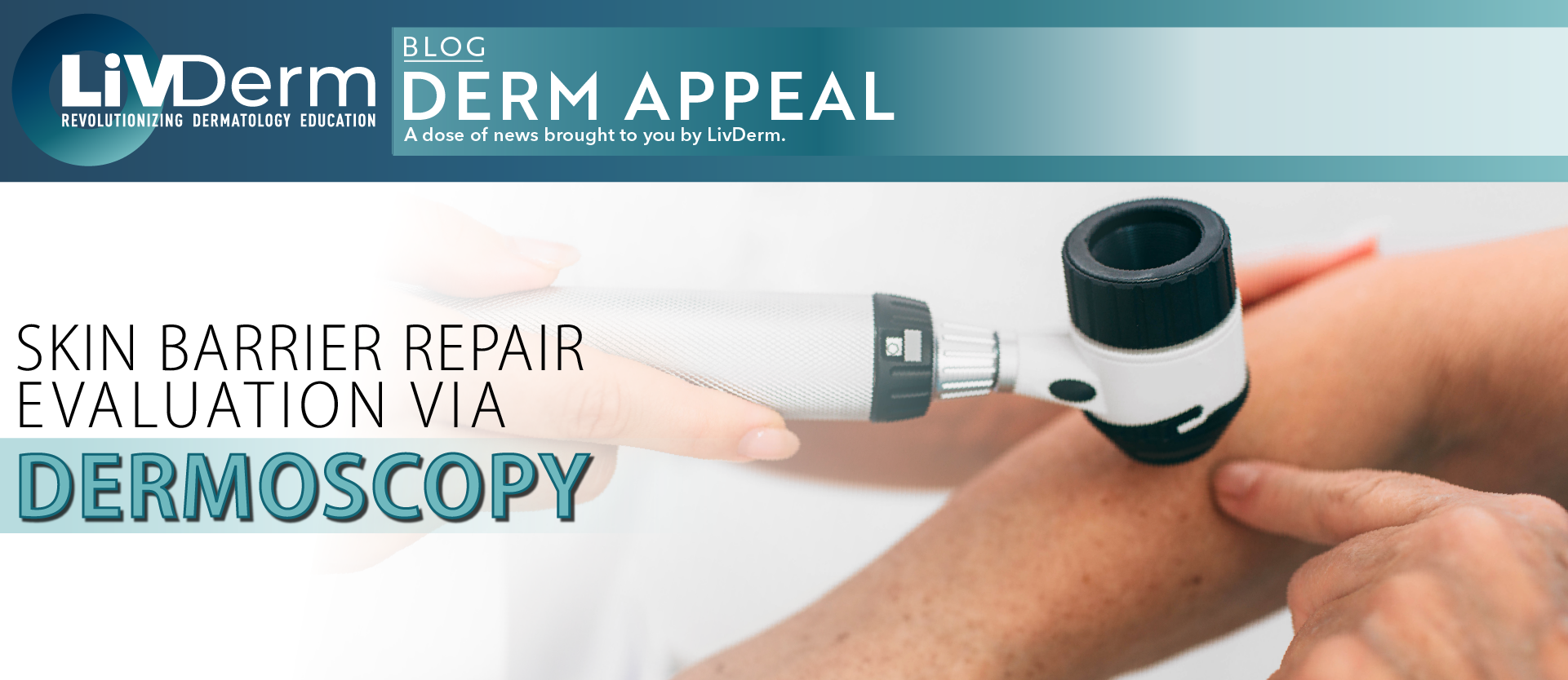
Although primarily used for the assessment of skin lesions and other dermatologic disorders, dermoscopy holds promise as a potential tool for investigating skin barrier integrity and function. Recent study results published in the Journal of Cosmetic Dermatology reveal that the dermoscopy method proved to be an effective evaluation technique of the skin barrier repair process after damage.
Skin Barrier Damage and Repair
In the preliminary study, researchers at a major hospital in Guangzhou, China evaluated 25 healthy patients (mean age of 36.5 years) with normal forearm skin to investigate cutaneous changes under the dermoscope and to explore skin physiological indexes in the damage and repair process.
Trial participants underwent repeated tape-stripping of the skin to remove the stratum corneum followed by examination with the dermoscope in order to observe the process of skin repair.
Patients were divided into three groups based on the number of tape stripping repetitions at 30, 35, and 40 times for group A, group B, and group C, respectively.
Dermoscopy Observations
In group A, immediately after 30 tape stripping repetitions, a small amount of cuticle cells was detected via dermoscopy. In group B, following 35 repetitions, dermoscopic images revealed no cuticle cell residue and instead showed “blurry vessels.” Finally, in group C, branching vessels with no cuticle cell residue were found immediately after stripping.
After 3 days, no vessels were visible in any of the groups. At day 7, keratin was most apparent in group C compared to other groups. During the skin repair process, scab formation was detected by day 14 for group A, day 7 for group B, and day 3 for group C.
Researchers noted that mean TEWL measures were significantly different between groups A and B immediately after stripping, with group B experiencing more TEWL. Groups B and C did not differ in TEWL values at any point.
Stratifying against skin surface hydration, participants with higher hydration levels had unique visual dermoscopy characteristics. In particular, patients with greater skin surface hydration had no cuticle cell residue immediately after stripping and “blurry vessels” were visually apparent. Micro-scabs developed by day 7 in the group with lower hydration and day 3 in the group with higher hydration.
The researcher’s findings suggest dermoscopy may provide clinicians with useful information concerning the skin barrier repair process in patients with healthy skin as physiological changes could be clearly observed using the tool.
“By combining dermoscopy and skin indexes assessing technologies, the skin barrier integrity and function [can] be observed and evaluated more accurately and precisely,” investigators concluded. Nonetheless, further research is needed to determine whether the dermoscope can consistently deliver accurate results in a larger and more ethnically diverse study population before it can be applied in the clinical setting.
