Melanoma remains one of the most aggressive forms of skin cancer, but when detected early, it is highly treatable. As a dermatology professional, your front-line interactions with patients place you in a key position to support early detection and referral, particularly for individuals with an elevated risk of melanoma.
New tools like total body photography (TBP) are transforming how clinicians monitor high-risk patients and detect suspicious lesions. Understanding this technology and your role in the referral pipeline can improve outcomes and enhance the level of care your practice delivers.
What Is Total Body Photography?
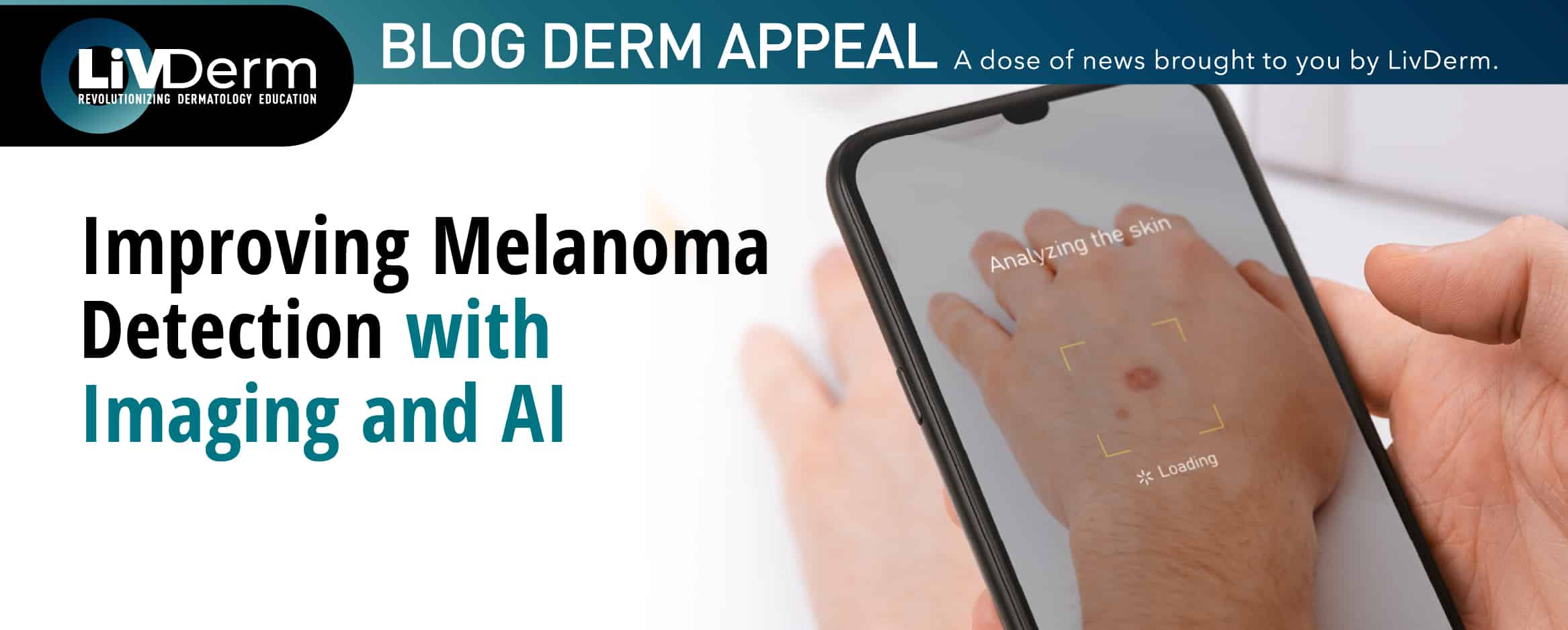
Total body photography is a noninvasive imaging method used to create a comprehensive visual record of a patient’s skin. The process involves capturing full-body images followed by high-resolution dermoscopic imaging of existing moles or lesions.
This system is designed to track changes over time, detecting new lesions or shifts in existing ones that could signal melanoma. Nebraska Medicine, for example, utilizes the FotoFinder system, which provides:
- 100x magnification beneath the skin surface
- AI-assisted analysis of suspicious spots
- Automated tracking of mole evolution
- Cloud-based image storage linked to patient records
AI as a Diagnostic Ally in Melanoma Detection
While artificial intelligence (AI) is not a substitute for clinical judgment, it plays a growing role in supporting diagnostic accuracy. For high-risk patients with many lesions or irregular patterns, AI enables a level of surveillance that would be difficult to maintain manually.
As a professional in the dermatologic space, it’s important to understand how AI supports, not replaces, the provider-patient relationship. You can help educate patients on its role and reassure them that decisions about biopsies or treatment still involve direct clinical oversight.
Who Should Be Referred for TBP?
Total body photography isn’t recommended for everyone. It’s a targeted tool best reserved for patients with known risk factors for melanoma, such as:
- A personal history of melanoma
- A strong family history of melanoma
- Genetic syndromes that increase melanoma risk
- Numerous or atypical nevi
- A provider-determined high-risk classification
Educating patients about their risk level and when additional imaging may be appropriate strengthens your value as a preventative care advocate.
How You Can Integrate TBP Awareness into Your Practice
While you may not offer TBP in-house, incorporating basic melanoma education into regular consultations is both proactive and professional. This includes:
- Performing regular skin assessments during injectable or facial appointments
- Encouraging annual skin checks
- Discussing TBP as a proactive surveillance option for high-risk individuals
- Identifying concerning lesions and referring for evaluation
These efforts demonstrate your commitment to whole-patient care, which ultimately strengthens client relationships and trust.


